40 cow eye labeled diagram
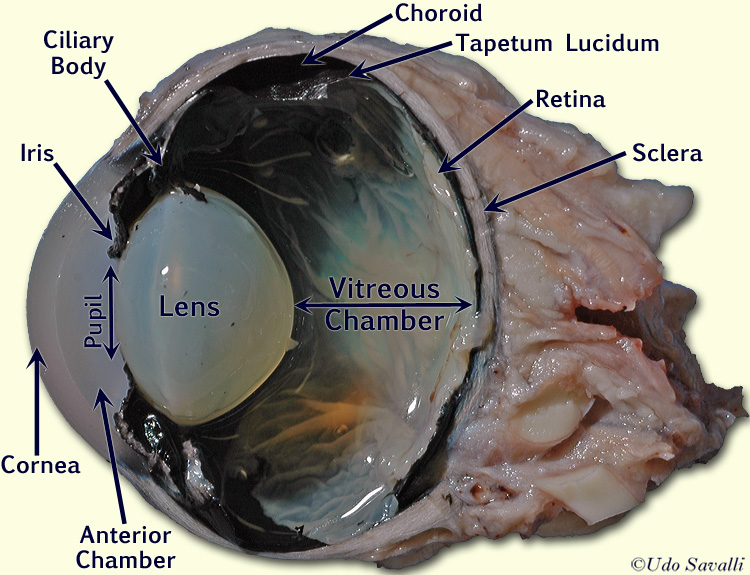
DOC Cow Eye Dissection - Western Michigan University For this lab exercise, we will examine the internal anatomy of the cow eye. Cow eyes are relatively inexpensive, and are particularly useful because of their size. Moreover, the basic structures are similar to those in the primate eye. Most diagrams of the eye show a cross section to illustrate the relative locations of all the important ... PDF Cow Eye Dissection Guide - Central Bucks School District DISSECTION OF THE COW EYE Please make sure to wear gloves and safety glasses when you are dissecting, and make sure to clean up thoroughly after the lab. Also, the cow eyes can be rather slippery, so use caution when handling and cutting them. You will need a scalpel and forceps. 1. First, identify the most external structures of the eye.
Diagram of Beef Cuts - Tyner Pond Farm Here's our Cow Map, a diagram of all the key sections of a cow and what cuts of beef come from each area. Key Sections of a Cow Here are the major sections of a butchered cow along with the cuts of beef that come from them. Brisket - brisket flat cut
Cow eye labeled diagram
Cow Eye Dissection Guide - Google Slides Cow Eye Use the point of a scissors or a scalpel to make an incision through the layers of the eye capsule (similar to figure 1); there are three layers from the exterior: sclera, whitish/grey,... Cow Eye Dissection & Anatomy Project | HST Learning Center Cow Eye Dissection: Internal Anatomy 1. Place the cow's eye on a dissecting tray. The eye most likely has a thick covering of fat and muscle tissue. Carefully cut away the fat and the muscle. As you get closer to the actual eyeball, you may notice muscles that are attached directly to the sclera and along the optic nerve. Cow Eye Dissection - The Biology Corner The cow eye is a fantastic specimen for students of all ages to dissect. ... I often leave eye models out during the lab to show comparisons between the cow and the human eye. Plus, I've found that my anatomy students have trouble matching parts on models to the real thing. I also leave diagrams on lab tables to help locate structures. Common ...
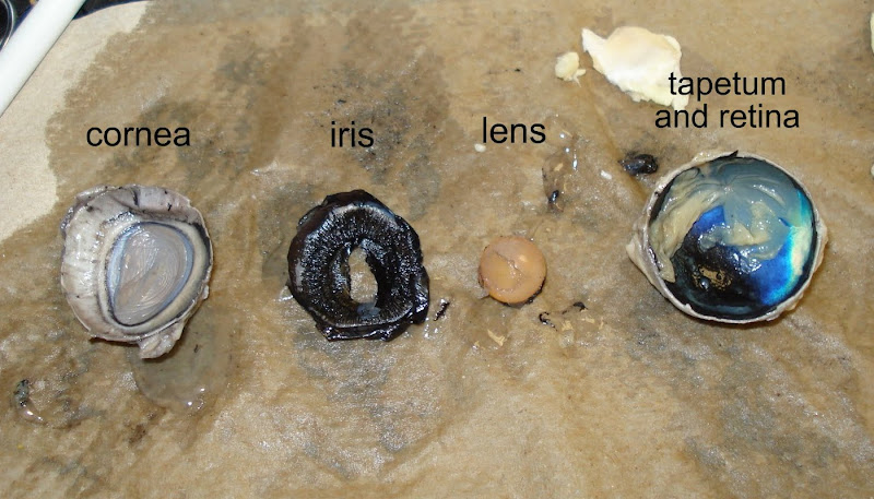
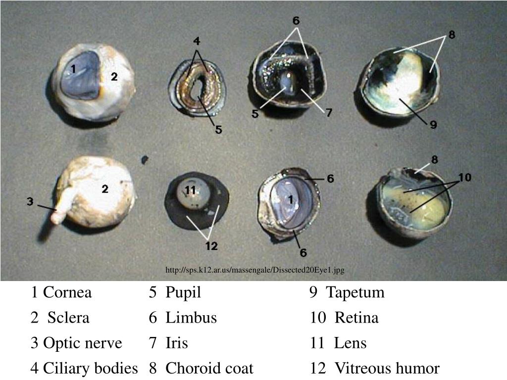
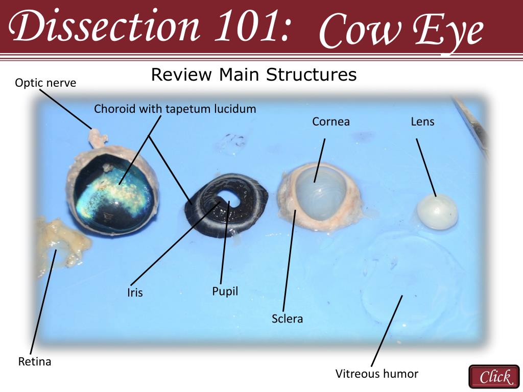
Cow eye labeled diagram. Cow Eye Dissection Diagram | Quizlet Located in the back of the eye, contains the rods and cones. pupil This structure of the eye can be dilated or constricted to control the amount of light entering the eye. lens The transparent structure behind the pupil that changes shape to help focus images on the retina. optic disc Location of the retina where there are no rods or cones. Cow's Eye Diagram Quiz - PurposeGames.com About this Quiz This is an online quiz called Cow's Eye Diagram There is a printable worksheet available for download here so you can take the quiz with pen and paper. From the quiz author Label the parts of the eye. This quiz has tags. Click on the tags below to find other quizzes on the same subject. biology eye light cow diagram optics Cow Eye Dissection | Carolina.com Students explore the external and internal anatomy, learning how structures work together to create images from incoming light. A preserved cow eye dissection can be carried out in 1-2 class periods and only requires basic dissecting instruments. Explore the internal and external anatomy of the cow eye using the procedural steps below. Cow Eye Dissection - The Biology Corner 1. Examine the outside of the eye. You should be able to find the sclera, or the whites of the eye. This tough, outer covering of the eyeball has fat and muscle attached to it. 2. Locate the covering over the front of the eye, the cornea. When the cow was alive, the cornea was clear. In your cow's eye, the cornea may be cloudy or blue in color.
Cow's Eye Dissection - Eye diagram - Exploratorium A cow's iris is brown. many colors, including brown, blue, green, and gray. A clear fluid that helps the cornea keep its rounded shape. The pupil is the dark circle in the center of your iris. It's a hole that Your pupil is round. is oval. A tough, Light bends as it passes through the cornea. Cow Anatomy - External Body Parts and Internal Organs with Labeled Diagram I will try to show you all of the body parts of a cow in labeled diagrams. The external body parts from the head region of a cow - in this head region, you might identify the mouth, lip, cheek, chin, muzzle, forehead, poll, ear, eye, nostril, and other. Cow Eye Labeled Diagram - ClipArt Best 31 cow eye labeled diagram. Free cliparts that you can download to you computer and use in your designs. Cow Eye Dissection - YouTube About Press Copyright Contact us Creators Advertise Developers Terms Privacy Policy & Safety How YouTube works Test new features Press Copyright Contact us Creators ...
10 Cow eye ideas | cow eyes, eye anatomy, anatomy and ... - Pinterest Sep 18, 2017 - Explore Allison Yorgey's board "cow eye" on Pinterest. See more ideas about cow eyes, eye anatomy, anatomy and physiology. Cow Skull Anatomy - The Place to Learn Veterinary Anatomy Online Labeled cow skull anatomy. Again, in this part, I will show you all the bones with a labeled cow skull anatomy diagram. You may also get help from the cow skull bones identification video. If you need more labeled diagrams or labeled cow skull pictures, you may join with anatomy learners on social media. Frequently asked questions on cow skull. Cow's Eye Dissection - Eye diagram - Exploratorium Learn how to dissect a cow's eye in your classroom. This resource includes: a step-by-step, hints and tips, a cow eye primer, and a glossary of terms. Cow's Eye Dissection - Eye diagram PDF Cow Eye Dissection: Examining Structure and Function These muscles allow the eye to move in different directions so that the animal can see more of its surroundings without turning its head. 3. Trim the fat and muscle from around the eye. Be careful not to cut the optic nerveon the back of the eye. 4. Using scissors or a scalpel, carefully cut the eye in half.
Cow Eye Dissection
General anatomy of the bull and the cow - IMAIOS Bovine anatomy - Illustrated atlas. This module of vet-Anatomy provides the basics on the anatomy of the bull for students of veterinary medicine. This veterinary anatomical atlas includes 27 scientific illustrations with a selection of labelled structures to understand and discover animal anatomy (skeleton, bones, muscles, joints and viscera).
PPT - COW EYE DISSECTION PowerPoint Presentation, free download - ID ...
[Solved] How to label a cow eye dissection? | Course Hero You can now label your diagram accordingly. There are a few different ways that you can label a cow eye dissection. One way is to use a permanent marker to label the different parts of the eye. Another way is to use a piece of tape and write the labels on the tape. You can also use labels that are made specifically for eye dissections.

Smith, K / Anatomy and Physiology Activities
Cow Eye Parts Labeled - All About Cow Photos Cow Eye Labeled Clipart Best Development Anatomy And Physiology Of The Eye Word Perspective Es From Latin Per Through Specere Look Anatomy Of The Eye Sheep Eye Dissection Lab Lab 15 Cow Eye Dissection Flashcards Quizlet Cow S Eye Diagram Quiz Ppt Cow Eye Dissection Powerpoint Ation Id 3482425 Cow Eye Dissection Anatomy Hst Learning Center

DISSECTING A COW'S EYE | business-strategy-bl
Diagram Of Cow High Resolution Stock Photography and Images - Alamy Find the perfect diagram of cow stock photo. Huge collection, amazing choice, 100+ million high quality, affordable RF and RM images. No need to register, buy now!
LAS Assignment 7 - Eye Enucleation
Cow Eye Dissection & Parts of the Eye Diagram | Quizlet cornea Clear, outer layer of the front of the eye. sclera White, outermost layer of the eye. Helps maintain shape and gives attachment to muscles. photoreceptors The cells in the retina that respond to light (rods and cones) rods Photoreceptor cells in the eye that detect black, white, and gray cones Photoreceptor cells in the eye that detect color
PPT - Reasons to Use the Dissection Video and Accompanying PowerPoint ...
Cow Eye Dissection Teaching Resources | Teachers Pay Teachers Cow Eye Dissection by CrazyScienceLady 49 $5.00 PDF Cow Eye Dissection: Directions and questions for dissecting a cow eye. Also includes a diagram of the eye, fun facts, an activity on how to find your blind spot and all answer keys. Appropriate for grades 4-7, I run this lab for each of the 4th grade classes at my school. It's a HUGE hit!!!

Cow Eye Dissection Worksheet | Cow eyes, Dissection, Frog dissection ...
Anatomy Of Eye Worksheets: Worksheets Cow Eye Dissection Worksheet ... Anatomy Of Eye Worksheets: Worksheets Cow Eye Dissection Worksheet Answers Laurenpsyk Free , ...
Post a Comment for "40 cow eye labeled diagram"